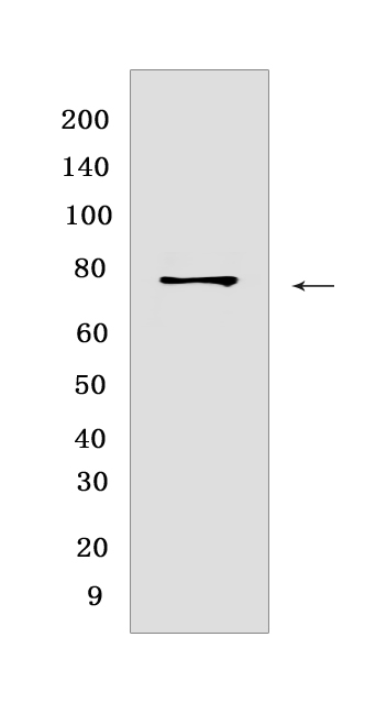
P-RIP (S166) Rabbit mAb [1Y74]Cat NO.: A86074
Western blot(SDS PAGE) analysis of extracts from HT-29 cells treated with Z-VAD (20 μM added 30 min prior to other compounds) Human TNF-α (20 ng/ml 7 hr) and SM-164 (100 nM 7 hr).Using P-RIP (S166) Rabbit mAb [1Y74] at dilution of 1:1000 incubated at 4℃ over night.
Product information
Protein names :RIPK1,RIP,RIP1,RIPK1_HUMAN,Receptor-interacting serine/threonine-protein kinase 1
UniProtID :Q13546
MASS(da) :75,931
MW(kDa) :78-82 kDa
Form :Liquid
Purification :Protein A purification
Host :Rabbit
Isotype :IgG
sensitivity :Endogenous
Reactivity :Human
- ApplicationDilution
- 免疫印迹(WB)1:1000-2000
- The optimal dilutions should be determined by the end user
Specificity :Antibody is produced by immunizing animals with a synthetic peptide at the sequence of Human Phospho-RIP (Ser166)
Storage :Antibody store in 10 mM PBS, 0.5mg/ml BSA, 50% glycerol. Shipped at 4°C. Store at-20°C or -80°C. Products are valid for one natural year of receipt.Avoid repeated freeze / thaw cycles.
WB Positive detected :HT-29 cells treated with Z-VAD (20 μM added 30 min prior to other compounds) Human TNF-α (20 ng/ml 7 hr) and SM-164 (100 nM 7 hr)
Function : Serine-threonine kinase which is a key regulator of TNF-mediated apoptosis, necroptosis and inflammatory pathways (PubMed:32657447, PubMed:31827280, PubMed:31827281). Exhibits kinase activity-dependent functions that regulate cell death and kinase-independent scaffold functions regulating inflammatory signaling and cell survival (PubMed:11101870, PubMed:19524512, PubMed:19524513, PubMed:29440439, PubMed:30988283). Has kinase-independent scaffold functions: upon binding of TNF to TNFR1, RIPK1 is recruited to the TNF-R1 signaling complex (TNF-RSC also known as complex I) where it acts as a scaffold protein promoting cell survival, in part, by activating the canonical NF-kappa-B pathway (By similarity). Kinase activity is essential to regulate necroptosis and apoptosis, two parallel forms of cell death: upon activation of its protein kinase activity, regulates assembly of two death-inducing complexes, namely complex IIa (RIPK1-FADD-CASP8), which drives apoptosis, and the complex IIb (RIPK1-RIPK3-MLKL), which drives necroptosis (By similarity). RIPK1 is required to limit CASP8-dependent TNFR1-induced apoptosis (By similarity). In normal conditions, RIPK1 acts as an inhibitor of RIPK3-dependent necroptosis, a process mediated by RIPK3 component of complex IIb, which catalyzes phosphorylation of MLKL upon induction by ZBP1 (PubMed:19524512, PubMed:19524513, PubMed:29440439, PubMed:30988283). Inhibits RIPK3-mediated necroptosis via FADD-mediated recruitment of CASP8, which cleaves RIPK1 and limits TNF-induced necroptosis (PubMed:19524512, PubMed:19524513, PubMed:29440439, PubMed:30988283). Required to inhibit apoptosis and necroptosis during embryonic development: acts by preventing the interaction of TRADD with FADD thereby limiting aberrant activation of CASP8 (By similarity). In addition to apoptosis and necroptosis, also involved in inflammatory response by promoting transcriptional production of pro-inflammatory cytokines, such as interleukin-6 (IL6) (PubMed:31827280, PubMed:31827281). Phosphorylates RIPK3: RIPK1 and RIPK3 undergo reciprocal auto- and trans-phosphorylation (PubMed:19524513). Phosphorylates DAB2IP at 'Ser-728' in a TNF-alpha-dependent manner, and thereby activates the MAP3K5-JNK apoptotic cascade (PubMed:17389591, PubMed:15310755). Required for ZBP1-induced NF-kappa-B activation in response to DNA damage (By similarity)..
Subcellular locationi :Cytoplasm. Cell membrane.
IMPORTANT: For western blots, incubate membrane with diluted primary antibody in 1% w/v BSA, 1X TBST at 4°C overnight.


