MICA Rabbit mAb [JjgS]Cat NO.: A72764
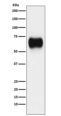
Western blot(SDS PAGE) analysis of extracts from A431 cell lysate.Using MICA Rabbit mAb [JjgS]at dilution of 1:1000 incubated at 4℃ over night.
Product information
Protein names :PERB11.1; MICA;
UniProtID :Q29983
MASS(da) :42,915
MW(kDa) :42-70kDa
Form :Liquid
Purification :Affinity-chromatography
Host :Rabbit
Isotype : IgG
sensitivity :Endogenous
Reactivity :Human
- ApplicationDilution
- 免疫印迹(WB)1:1000-2000
- The optimal dilutions should be determined by the end user
Specificity :Antibody is produced by immunizing animals with A synthesized peptide derived from human MICA
Storage :Antibody store in 10 mM PBS, 0.5mg/ml BSA, 50% glycerol. Shipped at 4°C. Store at-20°C or -80°C. Products are valid for one natural year of receipt.Avoid repeated freeze / thaw cycles.
WB Positive detected :A431 cell lysate.
Function : Seems to have no role in antigen presentation. Acts as a stress-induced self-antigen that is recognized by gamma delta T-cells. Ligand for the KLRK1/NKG2D receptor. Binding to KLRK1 leads to cell lysis..
Tissue specificity :Widely expressed with the exception of the central nervous system where it is absent. Expressed predominantly in gastric epithelium and also in monocytes, keratinocytes, endothelial cells, fibroblasts and in the outer layer of Hassal's corpuscles within the medulla of normal thymus. In skin, expressed mainly in the keratin layers, basal cells, ducts and follicles. Also expressed in many, but not all, epithelial tumors of lung, breast, kidney, ovary, prostate and colon. In thyomas, overexpressed in cortical and medullar epithelial cells. Tumors expressing MICA display increased levels of gamma delta T-cells..
Subcellular locationi :Cell membrane,Single-pass type I membrane protein. Cytoplasm.
IMPORTANT: For western blots, incubate membrane with diluted primary antibody in 1% w/v BSA, 1X TBST at 4°C overnight.


