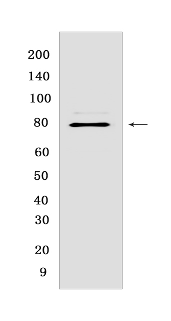
P-DRP1 (S616) Rabbit mAb [SFE8]Cat NO.: A19804
Western blot(SDS PAGE) analysis of extracts from HeLa cells.Using P-DRP1 (S616) Rabbit mAb [SFE8] at dilution of 1:1000 incubated at 4℃ over night.
Product information
Protein names :DNM1L,DLP1,DRP1,DNM1L_HUMAN,Dynamin-1-like protein
UniProtID :O00429
MASS(da) :81,877
MW(kDa) :78-82 kDa
Form :Liquid
Purification :Protein A purification
Host :Rabbit
Isotype :IgG
sensitivity :Endogenous
Reactivity :Human
- ApplicationDilution
- 免疫印迹(WB)1:1000-2000
- The optimal dilutions should be determined by the end user
Specificity :Antibody is produced by immunizing animals with a synthetic peptide at the sequence of Human Phospho-DRP1 (Ser616)
Storage :Antibody store in 10 mM PBS, 0.5mg/ml BSA, 50% glycerol. Shipped at 4°C. Store at-20°C or -80°C. Products are valid for one natural year of receipt.Avoid repeated freeze / thaw cycles.
WB Positive detected :HeLa cells
Function : Functions in mitochondrial and peroxisomal division (PubMed:9570752, PubMed:9786947, PubMed:11514614, PubMed:12499366, PubMed:17301055, PubMed:17553808, PubMed:17460227, PubMed:18695047, PubMed:18838687, PubMed:19638400, PubMed:19411255, PubMed:19342591, PubMed:23921378, PubMed:23283981, PubMed:23530241, PubMed:29478834, PubMed:32484300, PubMed:27145208, PubMed:26992161, PubMed:27301544, PubMed:27328748). Mediates membrane fission through oligomerization into membrane-associated tubular structures that wrap around the scission site to constrict and sever the mitochondrial membrane through a GTP hydrolysis-dependent mechanism (PubMed:23530241, PubMed:23584531). The specific recruitment at scission sites is mediated by membrane receptors like MFF, MIEF1 and MIEF2 for mitochondrial membranes (PubMed:23921378, PubMed:23283981, PubMed:29899447). While the recruitment by the membrane receptors is GTP-dependent, the following hydrolysis of GTP induces the dissociation from the receptors and allows DNM1L filaments to curl into closed rings that are probably sufficient to sever a double membrane (PubMed:29899447). Acts downstream of PINK1 to promote mitochondrial fission in a PRKN-dependent manner (PubMed:32484300). Plays an important role in mitochondrial fission during mitosis (PubMed:19411255, PubMed:26992161, PubMed:27301544, PubMed:27328748). Through its function in mitochondrial division, ensures the survival of at least some types of postmitotic neurons, including Purkinje cells, by suppressing oxidative damage (By similarity). Required for normal brain development, including that of cerebellum (PubMed:17460227, PubMed:27145208, PubMed:26992161, PubMed:27301544, PubMed:27328748). Facilitates developmentally regulated apoptosis during neural tube formation (By similarity). Required for a normal rate of cytochrome c release and caspase activation during apoptosis,this requirement may depend upon the cell type and the physiological apoptotic cues (By similarity). Required for formation of endocytic vesicles (PubMed:9570752, PubMed:20688057, PubMed:23792689). Proposed to regulate synaptic vesicle membrane dynamics through association with BCL2L1 isoform Bcl-X(L) which stimulates its GTPase activity in synaptic vesicles,the function may require its recruitment by MFF to clathrin-containing vesicles (PubMed:17015472, PubMed:23792689). Required for programmed necrosis execution (PubMed:22265414). Rhythmic control of its activity following phosphorylation at Ser-637 is essential for the circadian control of mitochondrial ATP production (PubMed:29478834).., [Isoform 1]: Inhibits peroxisomal division when overexpressed.., [Isoform 4]: Inhibits peroxisomal division when overexpressed..
Tissue specificity :Ubiquitously expressed with highest levels found in skeletal muscles, heart, kidney and brain. Isoform 1 is brain-specific. Isoform 2 and isoform 3 are predominantly expressed in testis and skeletal muscles respectively. Isoform 4 is weakly expressed in brain, heart and kidney. Isoform 5 is dominantly expressed in liver, heart and kidney. Isoform 6 is expressed in neurons..
Subcellular locationi :Cytoplasm, cytosol. Golgi apparatus. Endomembrane system,Peripheral membrane protein. Mitochondrion outer membrane,Peripheral membrane protein. Peroxisome. Membrane, clathrin-coated pit. Cytoplasmic vesicle, secretory vesicle, synaptic vesicle membrane.
IMPORTANT: For western blots, incubate membrane with diluted primary antibody in 1% w/v BSA, 1X TBST at 4°C overnight.


