Phospho-FGF Receptor 1 (Tyr766) Rabbit mAb[19B2]Cat NO.: A38677
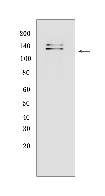
Western blot(SDS PAGE) analysis of extracts from COS cells overexpressing human FGFR1..Using Phospho-FGF Receptor 1 (Tyr766)Rabbit mAb IgG [19B2] at dilution of 1:1000 incubated at 4℃ over night.
Product information
Protein names :FGFR1,BFGFR,CEK,FGFBR,FLG,FLT2,HBGFR,FGFR1_HUMAN,Fibroblast growth factor receptor 1
UniProtID :P11362
MASS(da) :91,868
MW(kDa) : 120, 145
Form :Liquid
Purification :Protein A purification
Host :Rabbit
Isotype :IgG
sensitivity :Endogenous
Reactivity :Human
- ApplicationDilution
- 免疫印迹(WB)1:1000-2000
- The optimal dilutions should be determined by the end user
Specificity :Antibody is produced by immunizing animals with a synthetic peptide corresponding to residues surrounding Tyr766 of human FGF Receptor 1
Storage :Antibody store in 10 mM PBS, 0.5mg/ml BSA, 50% glycerol. Shipped at 4°C. Store at-20°C or -80°C. Products are valid for one natural year of receipt.Avoid repeated freeze / thaw cycles.
WB Positive detected :COS cells overexpressing human FGFR1.
Function : Tyrosine-protein kinase that acts as cell-surface receptor for fibroblast growth factors and plays an essential role in the regulation of embryonic development, cell proliferation, differentiation and migration. Required for normal mesoderm patterning and correct axial organization during embryonic development, normal skeletogenesis and normal development of the gonadotropin-releasing hormone (GnRH) neuronal system. Phosphorylates PLCG1, FRS2, GAB1 and SHB. Ligand binding leads to the activation of several signaling cascades. Activation of PLCG1 leads to the production of the cellular signaling molecules diacylglycerol and inositol 1,4,5-trisphosphate. Phosphorylation of FRS2 triggers recruitment of GRB2, GAB1, PIK3R1 and SOS1, and mediates activation of RAS, MAPK1/ERK2, MAPK3/ERK1 and the MAP kinase signaling pathway, as well as of the AKT1 signaling pathway. Promotes phosphorylation of SHC1, STAT1 and PTPN11/SHP2. In the nucleus, enhances RPS6KA1 and CREB1 activity and contributes to the regulation of transcription. FGFR1 signaling is down-regulated by IL17RD/SEF, and by FGFR1 ubiquitination, internalization and degradation..
Tissue specificity :Detected in astrocytoma, neuroblastoma and adrenal cortex cell lines. Some isoforms are detected in foreskin fibroblast cell lines, however isoform 17, isoform 18 and isoform 19 are not detected in these cells..
Subcellular locationi :Cell membrane,Single-pass type I membrane protein. Nucleus. Cytoplasm, cytosol. Cytoplasmic vesicle.
IMPORTANT: For western blots, incubate membrane with diluted primary antibody in 1% w/v BSA, 1X TBST at 4°C overnight.


