VEGFR1 Rabbit mAb[3HFA]Cat NO.: A79975
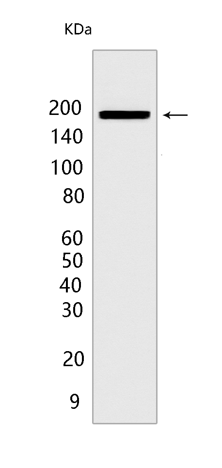
Western blot(SDS PAGE) analysis of extracts from Mouse brain tissue lysate.Using VEGFR1 Rabbit mAb IgG [3HFA] at dilution of 1:1000 incubated at 4℃ over night.
Product information
Protein names :FLT1,FLT,FRT,VEGFR1,VGFR1_HUMAN,Vascular endothelial growth factor receptor 1
UniProtID :P17948
MASS(da) :150,769
MW(kDa) :180kDa
Form :Liquid
Purification :Protein A purification
Host :Rabbit
Isotype :IgG
sensitivity :Endogenous
Reactivity :Mouse,Rat
- ApplicationDilution
- 免疫印迹(WB)1:1000-2000
- The optimal dilutions should be determined by the end user
Specificity :Antibody is produced by immunizing animals with a synthetic peptide of Mouse VEGFR1.
Storage :Antibody store in 10 mM PBS, 0.5mg/ml BSA, 50% glycerol. Shipped at 4°C. Store at-20°C or -80°C. Products are valid for one natural year of receipt.Avoid repeated freeze / thaw cycles.
WB Positive detected :Mouse brain tissue lysate
Function : Tyrosine-protein kinase that acts as a cell-surface receptor for VEGFA, VEGFB and PGF, and plays an essential role in the development of embryonic vasculature, the regulation of angiogenesis, cell survival, cell migration, macrophage function, chemotaxis, and cancer cell invasion. Acts as a positive regulator of postnatal retinal hyaloid vessel regression (Ref.11). May play an essential role as a negative regulator of embryonic angiogenesis by inhibiting excessive proliferation of endothelial cells. Can promote endothelial cell proliferation, survival and angiogenesis in adulthood. Its function in promoting cell proliferation seems to be cell-type specific. Promotes PGF-mediated proliferation of endothelial cells, proliferation of some types of cancer cells, but does not promote proliferation of normal fibroblasts (in vitro). Has very high affinity for VEGFA and relatively low protein kinase activity,may function as a negative regulator of VEGFA signaling by limiting the amount of free VEGFA and preventing its binding to KDR. Modulates KDR signaling by forming heterodimers with KDR. Ligand binding leads to the activation of several signaling cascades. Activation of PLCG leads to the production of the cellular signaling molecules diacylglycerol and inositol 1,4,5-trisphosphate and the activation of protein kinase C. Mediates phosphorylation of PIK3R1, the regulatory subunit of phosphatidylinositol 3-kinase, leading to activation of phosphatidylinositol kinase and the downstream signaling pathway. Mediates activation of MAPK1/ERK2, MAPK3/ERK1 and the MAP kinase signaling pathway, as well as of the AKT1 signaling pathway. Phosphorylates SRC and YES1, and may also phosphorylate CBL. Promotes phosphorylation of AKT1 at 'Ser-473'. Promotes phosphorylation of PTK2/FAK1 (PubMed:16685275).., [Isoform 1]: Phosphorylates PLCG.., [Isoform 2]: May function as decoy receptor for VEGFA.., [Isoform 3]: May function as decoy receptor for VEGFA.., [Isoform 4]: May function as decoy receptor for VEGFA.., [Isoform 7]: Has a truncated kinase domain,it increases phosphorylation of SRC at 'Tyr-418' by unknown means and promotes tumor cell invasion..
Tissue specificity :Detected in normal lung, but also in placenta, liver, kidney, heart and brain tissues. Specifically expressed in most of the vascular endothelial cells, and also expressed in peripheral blood monocytes. Isoform 2 is strongly expressed in placenta. Isoform 3 is expressed in corneal epithelial cells (at protein level). Isoform 3 is expressed in vascular smooth muscle cells (VSMC)..
Subcellular locationi :[Isoform 1]: Cell membrane,Single-pass type I membrane protein. Endosome.
IMPORTANT: For western blots, incubate membrane with diluted primary antibody in 1% w/v BSA, 1X TBST at 4°C overnight.


