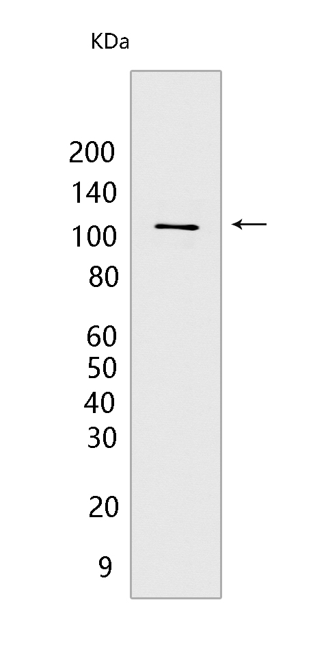
WWP1 Mouse mAb[Z028]Cat NO.: A84230
Western blot(SDS PAGE) analysis of extracts from MCF-7 cells.Using WWP1 Mouse mAb IgG [Z028] at dilution of 1:1000 incubated at 4℃ over night.
Product information
Protein names :WWP1,WWP1_HUMAN,NEDD4-like E3 ubiquitin-protein ligase WWP1
UniProtID :Q9H0M0
MASS(da) :105,202
MW(kDa) :105kda
Form :Liquid
Purification :Protein A purification
Host :Mouse
Isotype :IgG
sensitivity :Endogenous
Reactivity :Human
- ApplicationDilution
- 免疫印迹(WB)1:1000-2000
- The optimal dilutions should be determined by the end user
Specificity :Antibody is produced by immunizing animals with a synthetic peptide of human WWP1.
Storage :Antibody store in 10 mM PBS, 0.5mg/ml BSA, 50% glycerol. Shipped at 4°C. Store at-20°C or -80°C. Products are valid for one natural year of receipt.Avoid repeated freeze / thaw cycles.
WB Positive detected :MCF-7 cells
Function : E3 ubiquitin-protein ligase which accepts ubiquitin from an E2 ubiquitin-conjugating enzyme in the form of a thioester and then directly transfers the ubiquitin to targeted substrates. Ubiquitinates ERBB4 isoforms JM-A CYT-1 and JM-B CYT-1, KLF2, KLF5 and TP63 and promotes their proteasomal degradation. Ubiquitinates RNF11 without targeting it for degradation. Ubiquitinates and promotes degradation of TGFBR1,the ubiquitination is enhanced by SMAD7. Ubiquitinates SMAD6 and SMAD7. Ubiquitinates and promotes degradation of SMAD2 in response to TGF-beta signaling, which requires interaction with TGIF..
Tissue specificity :Detected in heart, placenta, pancreas, kidney, liver, skeletal muscle, bone marrow, fetal brain, and at much lower levels in adult brain and lung. Isoform 1 and isoform 5 predominate in all tissues tested, except in testis and bone marrow, where isoform 5 is expressed at much higher levels than isoform 1..
Subcellular locationi :Cytoplasm. Cell membrane,Peripheral membrane protein. Nucleus.
IMPORTANT: For western blots, incubate membrane with diluted primary antibody in 1% w/v BSA, 1X TBST at 4°C overnight.


