SIX2 Mouse mAb[61G7]Cat NO.: A91727
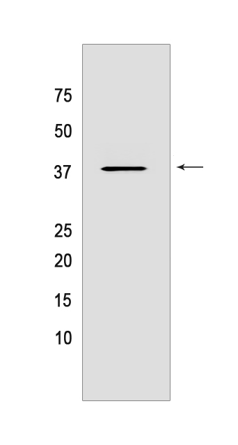
Western blot(SDS PAGE) analysis of extracts from HEK-293 cells.Using SIX2 Mouse mAb IgG [61G7] at dilution of 1:1000 incubated at 4℃ over night.
Product information
Protein names :SIX2,SIX2_HUMAN,Homeobox protein SIX2
UniProtID :Q9NPC8
MASS(da) :32,286
MW(kDa) :37kDa
Form :Liquid
Purification :Protein A purification
Host :Mouse
Isotype :IgG
sensitivity :Endogenous
Reactivity :Human,Mouse,Rat
- ApplicationDilution
- 免疫印迹(WB)1:1000-2000
- 免疫组化(IHC)1:100
- The optimal dilutions should be determined by the end user
Specificity :Antibody is produced by immunizing animals with a synthetic peptide of human SIX2.
Storage :Antibody store in 10 mM PBS, 0.5mg/ml BSA, 50% glycerol. Shipped at 4°C. Store at-20°C or -80°C. Products are valid for one natural year of receipt.Avoid repeated freeze / thaw cycles.
WB Positive detected :HEK-293 cells
Function : Transcription factor that plays an important role in the development of several organs, including kidney, skull and stomach. During kidney development, maintains cap mesenchyme multipotent nephron progenitor cells in an undifferentiated state by opposing the inductive signals emanating from the ureteric bud and cooperates with WNT9B to promote renewing progenitor cells proliferation. Acts through its interaction with TCF7L2 and OSR1 in a canonical Wnt signaling independent manner preventing transcription of differentiation genes in cap mesenchyme such as WNT4. Also acts independently of OSR1 to activate expression of many cap mesenchyme genes, including itself, GDNF and OSR1. During craniofacial development plays a role in growth and elongation of the cranial base through regulation of chondrocyte differentiation. During stomach organogenesis, controls pyloric sphincter formation and mucosal growth through regulation of a gene network including NKX2-5, BMPR1B, BMP4, SOX9 and GREM1. During branchial arch development, acts to mediate HOXA2 control over the insulin-like growth factor pathway. Also may be involved in limb tendon and ligament development (By similarity). Plays a role in cell proliferation and migration..
Tissue specificity :Strongly expressed in skeletal muscle. Expressed in Wilms' tumor and in the cap mesenchyme of fetal kidney (at protein level)..
Subcellular locationi :Nucleus.
IMPORTANT: For western blots, incubate membrane with diluted primary antibody in 1% w/v BSA, 1X TBST at 4°C overnight.


