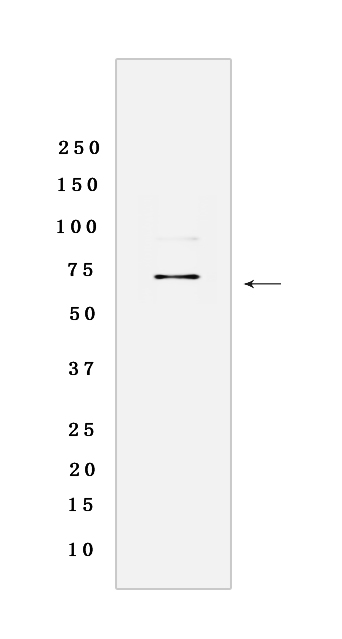
Ezrin Rabbit mAb [80RA]Cat NO.: A44074
Western blot analysis of extracts from HeLa cells lyastes.using Ezrin Rabbit mAb [80RA] at dilution of 1:1000 incubated at 4℃ over night
Product information
Protein names :EZR,VIL2,EZRI_HUMAN,Ezrin
UniProtID :P15311
MASS(da) :69,413
MW(kDa) :69 kDa
Form :Liquid
Purification :Protein A purification
Host :Rabbit
Isotype :IgG
sensitivity :Endogenous
Reactivity :Human,Mouse
- ApplicationDilution
- 免疫印迹(WB)1:1000-2000
- The optimal dilutions should be determined by the end user
Specificity :Antibody is produced by immunizing animals with a synthetic peptide of Human Ezrin.
Storage :Antibody store in 10 mM PBS, 0.5mg/ml BSA, 50% glycerol. Shipped at 4°C. Store at-20°C or -80°C. Products are valid for one natural year of receipt.Avoid repeated freeze / thaw cycles.
WB Positive detected :HeLa cells lyastes
Function : Probably involved in connections of major cytoskeletal structures to the plasma membrane. In epithelial cells, required for the formation of microvilli and membrane ruffles on the apical pole. Along with PLEKHG6, required for normal macropinocytosis..
Tissue specificity :Expressed in cerebral cortex, basal ganglia, hippocampus, hypophysis, and optic nerve. Weakly expressed in brain stem and diencephalon. Stronger expression was detected in gray matter of frontal lobe compared to white matter (at protein level). Component of the microvilli of intestinal epithelial cells. Preferentially expressed in astrocytes of hippocampus, frontal cortex, thalamus, parahippocampal cortex, amygdala, insula, and corpus callosum. Not detected in neurons in most tissues studied..
Subcellular locationi :Apical cell membrane,Peripheral membrane protein,Cytoplasmic side. Cell projection. Cell projection, microvillus membrane,Peripheral membrane protein,Cytoplasmic side. Cell projection, ruffle membrane,Peripheral membrane protein,Cytoplasmic side. Cytoplasm, cell cortex. Cytoplasm, cytoskeleton. Cell projection, microvillus.
IMPORTANT: For western blots, incubate membrane with diluted primary antibody in 1% w/v BSA, 1X TBST at 4°C overnight.


