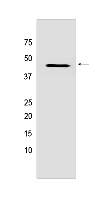
PELO Rabbit mAb [Z467]Cat NO.: A12066
Western blot analysis of extracts from HeLa cells lyastes.using PELO Rabbit mAb [Z467] at dilution of 1:1000 incubated at 4℃ over night
Product information
Protein names :PELO,CGI-17,PELO_HUMAN,Protein pelota homolog
UniProtID :Q9BRX2
MASS(da) :43,359
MW(kDa) :43 kDa
Form :Liquid
Purification :Protein A purification
Host :Rabbit
Isotype :IgG
sensitivity :Endogenous
Reactivity :Human
- ApplicationDilution
- 免疫印迹(WB)1:1000-2000
- 免疫组化(IHC)1:100,
- The optimal dilutions should be determined by the end user
Specificity :Antibody is produced by immunizing animals with a synthetic peptide of Human PELO.
Storage :Antibody store in 10 mM PBS, 0.5mg/ml BSA, 50% glycerol. Shipped at 4°C. Store at-20°C or -80°C. Products are valid for one natural year of receipt.Avoid repeated freeze / thaw cycles.
WB Positive detected :HeLa cells lyastes
Function : Cotranslational quality control factor involved in the No-Go Decay (NGD) pathway (PubMed:21448132, PubMed:29861391). In the presence of ABCE1 and HBS1L, is required for 48S complex formation from 80S ribosomes and dissociation of vacant 80S ribosomes (PubMed:21448132). Together with HBS1L and in presence of ABCE1, recognizes stalled ribosomes and promotes dissociation of elongation complexes assembled on non-stop mRNAs,this triggers endonucleolytic cleavage of the mRNA, a mechanism to release non-functional ribosomes and to degrade damaged mRNAs as part of the No-Go Decay (NGD) pathway (PubMed:21448132). As part of the PINK1-regulated signaling, upon mitochondrial damage is recruited to the ribosome/mRNA-ribonucleoprotein complex associated to mitochondrial outer membrane thereby enabling the recruitment of autophagy receptors and induction of mitophagy (PubMed:29861391)..
Tissue specificity :Ubiquitously expressed..
Subcellular locationi :Nucleus. Cytoplasm.
IMPORTANT: For western blots, incubate membrane with diluted primary antibody in 1% w/v BSA, 1X TBST at 4°C overnight.


