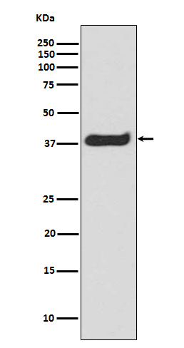
P-LAT (Y220) Rabbit mAb [M3Wf]Cat NO.: A38759
Western blot(SDS PAGE) analysis of extracts from Jurkat cell treated with CD3 lysate.Using P-LAT (Y220) Rabbit mAb [M3Wf]at dilution of 1:1000 incubated at 4℃ over night.
Product information
Protein names :36 kDa phospho-tyrosine adaptor protein; LAT1; lat; pp36
UniProtID :O43561
MASS(da) :27,930
MW(kDa) :38kDa
Form :Liquid
Purification :Affinity-chromatography
Host :Rabbit
Isotype : IgG
sensitivity :Endogenous
Reactivity :Human,Mouse,Rat
- ApplicationDilution
- 免疫印迹(WB)1:1000-2000
- 免疫组化(IHC)1:100
- 免疫荧光(ICC/IF)1:100
- The optimal dilutions should be determined by the end user
Specificity :Antibody is produced by immunizing animals with A synthesized peptide derived from human Phospho-LAT (Y220)
Storage :Antibody store in 10 mM PBS, 0.5mg/ml BSA, 50% glycerol. Shipped at 4°C. Store at-20°C or -80°C. Products are valid for one natural year of receipt.Avoid repeated freeze / thaw cycles.
WB Positive detected :Jurkat cell treated with CD3 lysate.
Function : Required for TCR (T-cell antigen receptor)- and pre-TCR-mediated signaling, both in mature T-cells and during their development. Involved in FCGR3 (low affinity immunoglobulin gamma Fc region receptor III)-mediated signaling in natural killer cells and FCER1 (high affinity immunoglobulin epsilon receptor)-mediated signaling in mast cells. Couples activation of these receptors and their associated kinases with distal intracellular events such as mobilization of intracellular calcium stores, PKC activation, MAPK activation or cytoskeletal reorganization through the recruitment of PLCG1, GRB2, GRAP2, and other signaling molecules..
Tissue specificity :Expressed in thymus, T-cells, NK cells, mast cells and, at lower levels, in spleen. Present in T-cells but not B-cells (at protein level)..
Subcellular locationi :Cell membrane,Single-pass type III membrane protein.
IMPORTANT: For western blots, incubate membrane with diluted primary antibody in 1% w/v BSA, 1X TBST at 4°C overnight.


